Alışveriş sepeti (0) Ürün Ürünler (0)
Sipariş listesinde ürün yok
Kargo Bedava! Kargo
0,00 TL Toplam
Ürün başarıyla alışveriş sepetinize eklendi
Miktarı
Toplam
Sepetinizde 0 ürün bulunmaktadır. Sepetinizde 1 ürün bulunmaktadır.
Toplam ürün:
Restoratif Diş Tedavisi
- Tıp Kitapları
- Acil Tıp
- Adli Tıp ve kriminoloji
- Aile Hekimliği
- Alerji ve İmmünoloji
- Anatomi Kitapları
- Anesteziyoloji ve Ağrı Kitapları
- Biyoloji ve Genetik Kitapları
- Biyomedikal Mühendisliği
- Biyokimya Kitapları
- Çocuk Cerrahisi
- Çocuk Sağlığı ve Hastalıkları Kitabı
- Çocuk ve Ergen Psikiyatrisi
- Dahiliye Kitapları
- Dermatoloji Kitapları
- Endokrinoloji Kitapları
- Farmakoloji Kitapları
- Fiziksel Tıp ve Rehabilitasyon
- Fizyoterapi, Rehabilitasyon ve Spor Hekimliği
- Fizyoloji Kitapları
- Gastroenteroloji Kitapları
- Geleneksel ve Tamamlayıcı Tıp
- Genel Cerrahi Kitapları
- Geriatri
- Göz Hastalıkları
- Göğüs Hastalıkları
- Göğüs Cerrahisi
- Halk Sağlığı Kitapları
- Hematoloji Kitapları
- Histoloji ve Embriyoloji Kitapları
- İnfeksiyon Hastalıkları
- Kadın Hastalıkları ve Doğum
- Kardiyoloji Kitapları
- Kalp Damar Cerrahisi
- Kulak Burun Boğaz Hastalıkları
- Mikrobiyoloji immunoloji Kİtapları
- Nöroşirürji
- Nefroloji
- Nöroloji
- Nükleer Tıp
- Onkoloji
- Ortopedi ve Travmatoloji
- Patoloji Kitapları
- Plastik Cerrahi
- Psikiyatri
- Radyasyon Onkoloji
- Radyoloji
- Romatoloji
- Sağlıklı Yaşıyoruz
- Spor Hekimliği
- Tıp Tarihi ve Tıp Etiği
- Tıbbı İstatistik Araştırma
- Tıp ve Sağlık Hukuku
- Tıbbi Laboratuvar Deney Bilimi
- USMLE & Board Review
- Uyku Tıbbı
- Üroloji
- Yoğun Bakım
- Diş Hekimliği Kitapları
- Eczacılık Kitapları
- Beslenme ve Diyet Kitapları
- Veteriner Hekimlik
- DUS Kitapları
- DUS Akademi Konu Kitapları Serisi
- DUS için Açıklamalı Deneme Sınavları Serisi
- DUS Spot Bilgiler Serisi
- Miadent Konu Kitapları Serisi
- Miadent Soru Kitapları Serisi
- DUS Çıkmış Soru Kitapları
- DUSDATA Şampiyonların Notu
- DUS Review Serisi
- DUSDATAMAX Soru Kitapları Serisi
- DUS Akademi Soru Kitapları Serisi
- Diğer Kitapları Serisi
- TUS Kitapları
- Çıkmış TUS Soru Kitapları
- 41 Deneme Serisi
- MEDOTOMY Serisi
- Tusmer
- Klinisyen Konu Kitapları Serisi
- Optimum Serisi
- Premium Serisi
- PRETUS Deneme Sınavları Serisi
- ProspekTUS Serisi
- Klinisyen Soru Kitapları Serisi
- Tusdata Ders Notları
- Tıbbi İngilizce
- Vaka Soruları Serisi
- Tüm Tus Soruları
- Hızlı Tekrar Serisi
- UTS Serisi
- KAMP ÖZEL NOTLARI
- Meditus Serisi
- YDUS Kitapları
- Hemşirelik ve Ebelik kitapları
- HEMŞİRELİK / halk sağlığı
- HEMŞİRELİK / Hemşirelik Esasları
- HEMŞİRELİK / İç Hastalıkları
- HEMŞİRELİK / Cerrahi Hastalıkları
- HEMŞİRELİK / Kadın hastalıkları ve doğum Ebelik
- HEMŞİRELİK / Ruh Sağlığı ve Hastalıkları
- HEMŞİRELİK / Hemşirelikte Eğitim
- HEMŞİRELİK / Çocuk Sağlığı ve Hastalıkları
- HEMŞİRELİK / Acil tıp hemşireliği
- SAĞLIK BİLİMLERİ
- Çocuk Gelişimi
- Sağlık Yöneticiliği
- Optisyenlik
- Odyoloji
- Saç Bakımı ve Güzellik Hizmetleri
- Anestezi Teknikerliği
- Tıbbi Dökümantasyon ve Sekreterlik
- Tıbbi Laboratuvar Teknisyenliği
- İş Sağlığı ve Güvenliği
- Ergoterapi
- Ağız ve Diş Sağlığı Teknisyenliği
- Dil ve Konuşma Terapisi
- İlk ve Acil Yardım Teknikeri (Paramedik)
- Radyoloji Teknisyenliği
- EĞİTİM BİLİMLERİ
- Değerler Eğitimi
- Eğitim Programları ve Öğretim
- Eğitim Psikolojisi
- Eğitim Yönetimi ve Denetimi
- Eğitimde Drama
- Eğitim Temelleri
- Eğitim Teknolojileri
- Okul Öncesi Eğitim
- Ortaokul Öğretmenliği
- Öğretmenlik Eğitimi Bölümleri
- Ölçme ve Değerlendirme
- Özel Eğitim
- Psikolojik Danışmanlık ve Rehberlik
- Sınıf Öğretmenliği
- Sınıf Yönetimi Etkili Öğretim
- Yabancı Dil Eğitimi
- İLETİŞİM
- İŞLETME
- İKTİSAT / EKONOMİ / MALİYE
- MİMARLIK - SANAT
- BİLİM TEKNİK
- MÜHENDİSLİK - TEKNİK
- FEN BİLİMLERİ
- ÇOCUK VE GENÇLİK KİTAPLARI
- BEŞERİ/SOSYAL BİLİMLER
- ÇEVRE ve YER BİLİMLERİ
- GIDA TARIM ve HAYVANCILIK
- BİYOMEDİKAL MÜHENDİSLİĞİ
- SEYAHAT TURİZM
- SOSYAL ÇALIŞMALAR
- SPOR BİLİMLERİ
- YÖNETİM - SİYASET - ULUSLARARASI İLİŞKİLER
- SINAVLAR HAZIRLIK
- ÖNERİLEN ÜRÜNLER
- Çok Satan Romanlar
- E-Kitaplar
- AYBAK
- Kırtasiye
- Yabancı Dil Eğitimi
- AYBAK 2025 Bahar
 Daha büyük görüntüle
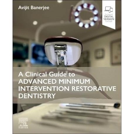
 A Clinical Guide to Advanced Minimum Intervention Restorative Dentistry
9780443109713
BU KİTAP İÇİN ÖN SİPARİŞ ALINMAKTADIR. TESLİM SÜRESİ 6 - 8 HAFTADIR. BİLGİ ALMAK İÇİN MAĞAZAMIZI ARAYINIZ
3 941,96 TL
3 153,57 TL
-20%
KDV Hariç: 3 153,57 TL
- Yorum Yaz
| As restorative dentistry shifts from a focus on core surgical procedures to the patient and their unique needs and values, this new book from acclaimed restorative dentistry expert Professor Avijit Banerjee is designed to support implementation of holistic patient care for long-term oral and dental health. The Guide to Advanced Minimum Intervention Restorative Dentistry describes the entire clinical journey through the minimum intervention oral healthcare delivery framework, with an emphasis on long term, risk-related, prevention-based care. It presents a blend of clinical and scientific evidence-based clinical protocols to guide the practitioner through the four domains of minimum intervention oral care - identifying disease, prevention / control, minimally invasive operative interventions, and review / re-assessment / active surveillance. Written in an engaging contemporary stylesss and easy to navigate, this important book is suitable for all members of the team, from undergraduates to experienced primary care practitioners and specialists alike. | ||
| Features: | ||
| ||
| Table Of Contents: | ||
| Foreword, v Preface, vi 1 Dental Hard Tissue Pathologies, 1 1.1 Dental Caries, 1 1.1.1 What Is It?, 1 1.1.2 Terminology, 1 1.1.3 Caries: The Process and the Lesion, 2 1.1.3.1 The Caries Process: Dental Plaque Biofilm, 2 1.1.3.2 The Carious Lesion: Dental Hard Tissues, 3 1.1.4 Aetiology of the Caries Process, 3 1.1.5 Speed and Severity of the Caries Process, 5 1.1.5.1 Definitions, 7 1.1.6 The Carious Lesion, 7 1.1.6.1 Within Enamel, 8 1.1.6.2 At the Enamel–Dentine Junction (EDJ)/Amelo-dentinal Junction, 10 1.1.6.3 Within Dentine, 10 1.1.7 Carious Pulp Exposure, 12 1.1.8 Dentine–Pulp Complex Reactions, 12 1.1.8.1 Translucent Dentine, 12 1.1.8.2 Tertiary Dentine, 13 1.1.8.3 Pulp Inflammation, 14 1.2 Toothwear, 15 1.3 Dental Trauma, 16 1.3.1 Aetiology, 17 1.4 Developmental Defects, 17 1.5 Causes of Tooth Discolouration, 17 SECTION 1 Minimum Intervention Oral Care (MIOC) – Clinical Domain 1 2 MIOC Domain 1: Identifying Clinical Problems, 27 2.1 Introduction: Minimum Intervention Oral Care and Minimally Invasive Dentistry, 27 2.2 Detection/Identification, 32 2.3 Taking a Verbal History, 33 2.4 Physical Examination, 33 2.4.1 General Examination, 33 2.4.2 Oral Examination, 33 2.4.3 Dental Charting, 36 2.4.4 Tooth Notation, 36 2.5 Caries Detection, 36 2.5.1 Caries Detection Indices, 38 2.5.2 Susceptible Surfaces, 41 2.5.3 Investigations, 44 2.5.3.1 Radiographs, 44 2.5.3.2 Pulp Sensibility Tests, 45 2.5.3.3 Percussion Tests, 49 2.5.4 Lesion Activity, 49 2.5.5 Diet Analysis, 51 2.5.6 Caries Detection Technologies, 54 2.6 Toothwear: Clinical Detection, 55 2.6.1 Targeted Verbal History, 55 2.6.2 Clinical Presentations of Toothwear, 55 2.6.3 Summary of the Clinical Manifestations of Toothwear, 57 2.7 Dental Trauma: Clinical Detection, 59 2.8 Developmental Defects: Clinical Detection, 60 3 MIOC Domain 1: Diagnosis, Prognosis and Personalised Care Planning, 65 3.1 Introduction, 65 3.1.1 Definitions, 65 3.2 Diagnosing Dental Pain, ‘Toothache’, 66 3.2.1 Acute Pulpitis, 66 3.2.2 Acute Periapical Periodontitis, 66 3.2.3 Acute Periapical Abscess, 69 3.2.4 Acute Periodontal (Lateral) Abscess, 69 3.2.5 Chronic Pulpitis, 69 3.2.6 Chronic Periapical Periodontitis, 69 3.2.7 Exposed Dentine Sensitivity, 70 3.2.8 Interproximal Food-Packing, 70 3.2.9 Cracked Cusp/Tooth Syndrome, 72 3.3 Caries Risk/Susceptibility Assessment (CRSA), 72 3.4 Diagnosing Toothwear, 75 3.5 Diagnosing Dental Trauma and Developmental Defects, 76 3.6 Prognostic Indicators, 76 3.7 Formulating a Risk-Related, Personalised Care Plan, 76 3.7.1 Why Is a Personalised Care Plan (PCP) Necessary?, 76 3.7.2 Structuring a Personalised Care Plan, 77 SECTION 2 Minimum Intervention Oral Care (MIOC) – Clinical Domain 2 4 MIOC Domain 2: Disease Control and Lesion Prevention, 83 4.1 Introduction, 83 4.1.1 Disease Control, 83 4.1.2 Lesion Prevention, 83 4.2 Caries Control and Lesion Prevention, 83 4.2.1 Categorising Caries Activity and Risk/ Susceptibility Status, 83 4.2.2 Behaviour Change/Modification: The COM-B Model, 84 4.2.3 Standard/Self Care: Low-Risk, Caries-Controlled, Disease-Inactive Patient, 87 4.2.3.1 Plaque Biofilm Control, 87 4.2.3.2 Fluoride, 90 4.2.3.3 Diet, 90 4.2.4 Active Care: High-Risk, Caries- Uncontrolled, Disease-Active Patient, 91 4.2.4.1 Decontamination Procedures, 91 4.2.4.2 Antimicrobial Agents, 91 4.2.4.3 Remineralisation, 92 4.3 Toothwear Control and Lesion Prevention, 99 4.3.1 Process, 99 4.3.2 Lesions, 99 SECTION 3 Minimum Intervention Oral Care (MIOC) – Clinical Domain 3 5 MIOC Domain 3: Minimally Invasive Operative Dentistry, 105 5.1 Clinical Management Guidelines, 106 5.2 The Dental Surgery, Clinic or Office, 107 5.2.1 Positioning the Clinician, Patient and Nurse, 107 5.2.2 Lighting, 108 5.2.3 Zoning, 108 5.3 Infection Prevention and Control/Personal Protective Equipment, 109 5.3.1 Decontamination and Sterilisation Procedures, 109 5.4 Patient Safety and Risk Management, 111 5.4.1 Management of Minor Injuries, 111 5.5 Dental Aesthetics, 112 5.5.1 Colour Perception, 113 5.5.2 Clinical Tips for Shade Selection, 114 5.5.3 MI Management of Tooth Discolouration, 116 5.6 Magnification, 118 5.6.1 Clinical Photography, 121 5.7 Moisture Control, 122 5.7.1 Why? 122 5.7.2 Techniques, 122 5.7.3 Rubber (Dental) Dam Placement: Practical Steps, 123 5.8 Instrumentation in Minimally Invasive Operative Dentistry, 128 5.8.1 Hand Instruments, 128 5.8.2 Rotary Instruments, 131 5.8.3 Using Hand/Rotary Instruments: Clinical Tips, 134 5.8.4 Dental Air-abrasion, 137 5.8.5 Chemo-mechanical Carious Tissue Removal, 137 5.8.6 Other Instrumentation Technologies, 138 5.9 “MI” Operative Management of the Cavitated Carious Lesion, 139 5.9.1 Rationale, 139 5.9.2 Minimally Invasive Dentistry (MID), 140 5.9.2.1 Assessing Clinical Cavitation of a Carious Lesion, 141 5.9.3 Enamel Preparation, 142 5.9.4 Carious Dentine Removal, 145 5.9.4.1 Peripheral Caries Management (EDJ), 146 5.9.4.2 Caries Overlying the Pulp, 147 5.9.4.3 Discriminating Between the Histological Zones of Carious Dentine, 149 5.9.4.4 ‘Stepwise Excavation’ and Atraumatic Restorative Technique, 149 5.9.5 Cavity Modifications, 151 5.10 Pulp Protection, 151 5.10.1 Rationale, 151 5.10.2 Terminology, 153 5.10.3 Materials, 153 5.11 Dental Matrices, 155 5.11.1 Clinical Tips, 156 5.11.2 Silicone Matrices, Stents and Splints, 156 5.11.3 ‘Stamp’ Matrix Technique, 157 5.12 Temporary/Provisional Restorations, 160 5.12.1 Definitions, 160 5.12.2 Clinical Tips, 160 5.13 Principles of Dental Occlusion, 160 5.13.1 Definitions, 160 5.13.2 Terminology, 161 5.13.3 Occlusal Registration Techniques, 161 5.13.4 Clinical Tips, 162 6 MIOC Domain 3: MI Operative Management of the Badly Broken Down Tooth, 166 6.1 Causes of Broken Down Teeth, 166 6.2 Clinical Assessment of Broken Down Teeth, 166 6.2.1 Why Restore the Broken Down (or any) Tooth?, 166 6.2.2 Is the Broken Down Tooth Restorable Clinically?, 167 6.3 Intra-coronal Core Restoration, 170 6.3.1 Direct Core Retention, 170 6.3.2 C-Factor, 170 6.4 Clinical Operative Tips, 171 6.5 Design Principles for Indirect Restorations, 172 6.5.1 Design Features, 173 7 MIOC Domain 3: Restorative Materials Used in Minimally Invasive Operative Dentistry, 176 7.1 Introduction, 177 7.2 Dental Resin Composites, 177 7.2.1 History, 177 7.2.2 Chemistry, 177 7.2.2.1 Resin Matrix, 177 7.2.2.2 Filler Particles, 178 7.2.2.3 Other Chemical Constituents, 180 7.2.2.4 Polymerisation Reaction, 181 7.2.3 The Tooth–Resin Composite Interface, 182 7.2.3.1 The Resin Composite–Enamel Interface, 183 7.2.3.2 Resin Composite – Dentine Interface: Dentine Bonding Agents/Dental Adhesives, 184 7.2.4 Classification of Dental Adhesives, 186 7.2.4.1 Type 1: Three-Bottle or Three-Step Bonding Systems, 186 7.2.4.2 Type 2: Two-Bottle or Two-Step, ‘Total Etch’ or ‘Etch and Rinse’ Adhesives, 187 7.2.4.3 Type 3: Weaker Self-Etching Primers, 187 7.2.4.4 Types 4 and 5: Self-Etching All-in- One/Universal Systems, 189 7.2.5 Clinical Issues With Dentine Bonding Agents/Dental Adhesives, 189 7.2.6 Developments, 190 7.2.6.1 Reducing Shrinkage Stress and Strain, 190 7.2.6.2 Dentine Bonding Agents, 190 7.2.6.3 ‘Self-Adhesive Composite’, 190 7.3 Glass-Ionomer Cement, 190 7.3.1 History, 190 7.3.2 Chemistry, 190 7.3.3 The Tooth–GIC Interface, 191 7.3.3.1 Enamel, 191 7.3.3.2 Dentine–Collagen, 191 7.3.3.3 Dentine–Tubules, 191 7.3.3.4 Surface Conditioning, 191 7.3.4 GIC Surface Protection , 191 7.3.5 Clinical Uses of GIC Relating to Its Properties, 194 7.3.6 Developments, 195 7.4 Resin-Modified Glass-Ionomer Cement and Polyacid-Modified Resin Composite (Compomer), 195 7.4.1 Chemistry, 196 7.4.1.1 RM-GICs, 196 7.4.1.2 Polyacid-Modified Composites, 196 7.4.2 Clinical Indications, 196 7.5 Dental Amalgam, 197 7.5.1 Chemistry, 197 7.5.2 Physical Properties, 198 7.5.3 Bonded and Sealed Amalgams, 198 7.5.3.1 Bonded Amalgams, 199 7.5.3.2 Sealed Amalgams, 199 7.5.4 Contemporary Indications for the Use of Dental Amalgam, 199 7.6 Temporary and Provisional (Intermediate) Restorative Materials, 199 7.6.1 Characteristics, 199 7.6.2 Chemistry, 200 7.7 Calcium Silicate Cements, 200 7.7.1 History, 200 7.7.2 Chemistry and Interactions With the Tooth, 200 7.7.3 Clinical Applications, 201 7.8 Materials and Techniques for Restoring the Endodontically Treated Tooth, 203 7.8.1 Root Canal Posts, 203 7.8.2 Root Canal Post Cementation, 203 8 MIOC Domain 3: Minimally Invasive Operative Dentistry (MID), a Step-by-Step Clinical Guide, 206 8.1 Introduction, 206 8.1.1 Cavity/Restoration Classification, 206 8.1.2 Restoration Procedures, 207 8.2 Preventive Fissure Sealant, 208 8.3 Therapeutic Sealant Restoration/Preventive Resin Restoration: Type 3 Adhesive, 211 8.4 Posterior Occlusal Resin Composite Restoration (Class I): Type 3 Adhesive, 216 8.5 Posterior Proximal Restoration (Class II), 221 8.5.1 Type 2 Adhesive, ‘Moist Bonding’, 221 8.5.2 Type 3 Adhesive, 225 8.6 Class III Anterior Restoration: Type 2 Adhesive, 232 8.7 Anterior Incisal Edge/Labial Veneer Composite (Class IV): Type 3 Adhesive (With Enamel Pre-etch), 235 8.8 Buccal Cervical Resin Composite Restorations (Class V): Type 2 Adhesive, 240 8.9 Posterior ‘Bonded’ Amalgam Restoration, 243 8.10 ‘Nayyar Core’ Restoration, 246 8.11 Direct Fibre-Post/Resin Composite Core Restoration, 247 8.12 Dentine Bonding Agents/Adhesives: A Stepby- Step Practical Guide for Use, 248 8.13 Checking the Final Restoration, 249 8.14 Patient Instructions, 249 SECTION 4 Minimum Intervention Oral Care (MIOC) – Clinical Domain 4 9 MIOC Domain 4: Clinical Re-assessment, Recall and Maintenance of the Tooth-Restoration Complex, 253 9.1 Active Surveillance of the Patient/Course of Disease, 253 9.1.1 Risk-related Re-assessment, 253 9.1.2 Re-assessment Checklist, 254 9.1.3 Monitoring Toothwear, 254 9.2 Restoration Failure, 256 9.2.1 Aetiology, 256 9.2.2 Choice of Restorative Material, 257 9.2.3 How May Restorations Be Assessed?, 259 9.2.4 How Long Should Restorations Last?, 259 9.3 Tooth Failure, 264 9.4 Managing the Failing Tooth-Restoration Complex: The ‘5Rs’, 264 9.4.1 Dental Amalgam, 267 9.4.2 Resin Composites/GIC, 270 10 MIOC Implementation: Personalised Care Management Pathways, 273 10.1 MIOC Framework: Summary, 273 10.1.1 Phased Courses of Treatment, 273 10.2 Care Management Pathways: Clinical Scenarios, 277 10.2.1 How Would You Manage the Following High-Risk Adult?, 278 10.2.2 How Would You Manage the Following High-Risk Child?, 282 10.2.3 How Would You Manage the Following High-Risk Older Adult Patient?, 285 10.2.4 How Would You Manage the Following Low-Risk Adult Patient?, 288 Index, 290 | ||
| ISBN | 9780443109713 |
| Basım Yılı | 2024 |
| Basım Sayısı | 1 |
| Sayfa Sayısı | 308 |
| Kitap Dili | İngilizce |
| Yazar(lar) | Avijit Banerjee |
| BISAC | |
| DOI |

